The Influence of PVOH Concentration on the Structural Morphology
and Dimension of Electrospun Nanofibers
Erni Misran
1,2
, Basuki Wirdjosentono
1,3
, Nasrudin M. Noor
4
, Saharman Gea
1,3
, Mahyuni Harahap
1,3
and Suhut Alexander Situmorang
1,3
1
Cellulosic and Functional Materials Research Centre, Universitas Sumatera Utara, Medan, Indonesia
2
Department of Chemical Engineering, Faculty of Engineering, Universitas Sumatera Utara, Medan, Indonesia
3
Department of Chemistry, Faculty of Mathematics and Natural Science, Universitas Sumatera Utara, Medan, Indonesia
4
Deparment of Physics, Faculty of Mathematics and Natural Science, Universitas Sumatera Utara, Medan, Indonesia
Keywords: Electrospinning, morphology, polyvinyl alcohol, nanofibers
Abstract: The parameters applied in electrospinning process were the main factors to produce ultrafine nanofibers for
different target applications. In this study, the effect of polyvinyl alcohol (PVOH) concentration, from 12%
to 16% (wt) in distilled water, on the morphology and dimension of nanofibers was investigated. Scanning
electron microscopy (SEM) was used to analyze the morphology of PVOH electrospun nanofibers and
Image J analysis was used to calculate the dimension of fibers. From the results, 12% and 13% of PVOH
concentration produced bead shaped fibers. Meanwhile ultrafine nanofibers were produced from PVOH
concentration of 14%, 15%, and 16% (wt). The diameter of ultrafine fibers increased with the increase in
PVOH concentration.
1 INTRODUCTION
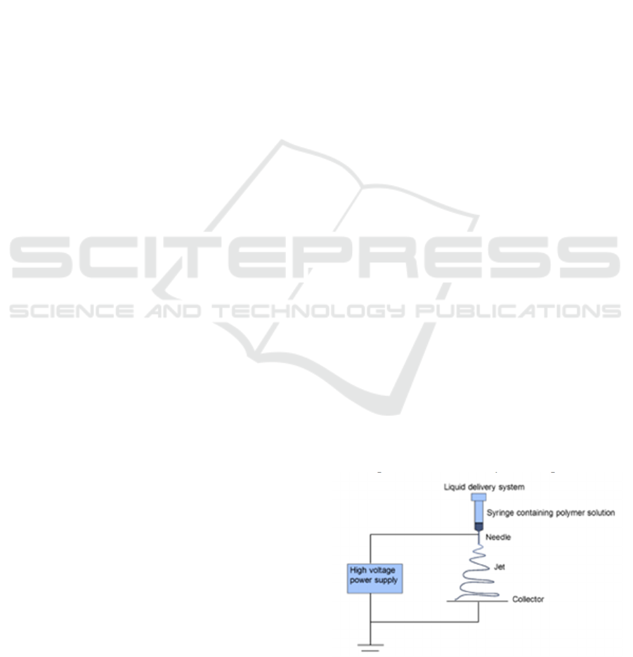
Electrospinning technique, with its simplicity setup
of syringe pump, needle, and collector only (Figure
1), has attracted significant interest among
researchers to fabricate fibers with diameters
ranging from microscale (10 m) to nanoscale
(<1000 nm) (Zhuo et al. 2008). Moreover, this
method provide higher surface area to volume ratio
compared to other methods, i.e. template synthesis,
self-assembly, phase separation and drawing
techniques (Abunahel et al. 2018). Electrospun
nanofibers have been applied to various applications
including tissue engineering, drug delivery system,
wound dressing, textile industry, cancer treatment
and the fabrication of new radiation shielding
material (Mirjalili and Zohoori, 2016).
Basically, the process of electrospinning uses
high voltage power supply, so there is a surface
charge at the end of the needle tip where the
polymer solution is held by its surface tension. Due
to the disturbance in the polymer surface, droplets
with spherical shapes called "Taylor zone" are
produced in the initiation of electrospinning process.
Then, a jet of polymer solution is ejected from the
tip of the needle to the collector. As the jet travels
through the air, the solvent evaporates and ultrafine
fibers are collected on the surface of the target (Teo
and Ramakrishna, 2006). The properties of spun
fibers can be controlled with three important
parameters; (i) the parameters of solution, which
include the concentration, molecular weight, surface
tension, conductivity, and volatility of polymer
solution; (ii) processing parameters, such as applied
voltage, liquid flow rate, and distance of the needle
to the collector; and (iii) processing environment
like temperature and humidity (Harahap 2018).
Figure 1 Electrospinning set up.
Misran, E., Wirdjosentono, B., Noor, N., Gea, S., Harahap, M. and Situmorang, S.
The Influence of PVOH Concentration on the Structural Morphology and Dimension of Electrospun Nanofibers.
DOI: 10.5220/0010613800002775
In Proceedings of the 1st International MIPAnet Conference on Science and Mathematics (IMC-SciMath 2019), pages 577-580
ISBN: 978-989-758-556-2
Copyright
c
2022 by SCITEPRESS – Science and Technology Publications, Lda. All rights reserved
577

There are more than 50 different polymers that
have been reported to have undergone successful
electrospinning. One of them is PVOH, a water-
soluble polymer, which can also be electrospun from
its aqueous solution phase (Lu et al. 2006). Many
studies have reported the influence of concentration
on the morphology of fibers, but there has not been
any literature data available reporting the diameters.
Hence, we aim to study the effect of PVOH
concentration to the morphology of electrospun
nanofibers, as well as their diameters.
2 EXPERIMENTAL
2.1 Materials
The material used in this study was PVOH powder,
Mw = 60,000 g/mole fully hydrolyzed, purchased
from Merck, Darmstadt, Germany. Before used,
PVOH was dried at 80 °C for 5 h in a vacuum oven
to remove the moisture content. Distilled water was
used as a solvent for PVOH.
2.2 Solution Preparation
PVOH solution with various concentration of 12%,
13%, 14%, 15%, and 16% (wt) was prepared under
reflux condition for 2 hours at 80 °C. After the
polymer was homogeneously mixed, the reaction
was put to stop and the solution was allowed to cool
with continuous stirring until it reached room
temperature. The solution was stored no more than
three days prior to use for electrospinning.
2.3 Electrospinning Process
The electrospinning process was carried out by
horizontal electrospinning (basic series
electrospinning unit Brand NLI, Nanolab
Instruments Sdn Bhd, Malaysia) at room
temperature. The condition was set-up as follow: (i)
disposable 18-G syringes; (ii) voltage was 15 kV;
(iii) polymer solution feed rate was 0.2 mL/hour;
(iv) needle tip-to-collector distance was 15 cm; and
(v) the speed of collector was 115 rpm. Ultrafine
fibers were collected on aluminum foils. The mat
fibers were dried in a vacuum oven at 40 °C for 3 h
to remove residual water and stored in a desiccator
containing silica gel.
2.3 Characterization
2.3.1 Scanning Electron Microscopy
The morphology of the samples was analyzed by
using a scanning electron microscopy (SEM) Hitachi
TM3030 (JEOL, Ltd., Tokyo, Japan). The sample
was coated with a thin layer of gold before analysis
to reduce charging. The diameter of the fibers was
calculated by using Image J software analysis.
3 RESULTS AND DISCUSSION
3.1 The Morphology
The concentration of the polymer solution is one of
the parameters controlled in the electrospinning
process. It has a big influence in the formation of
fibers. Low concentration (<1 Pa.s) has been
reported to form sprays instead of fibers. In this
condition, bead forms were also produced.
Meanwhile, fine fibers would be produced with
higher concentration of polymer solutions (Yang et
al. 2007).
In this study, PVOH solution dissolved in
distilled water was able to undergo electrospinning.
The fiber formation is illustrated in Figure 2. During
the process, there were no clogs at the tip of the
needle. Nonetheless, the morphology of PVOH
electrospun nanofibers were not the same for each
concentration used. SEM images for PVOH
nanofibers with concentration of 12%, 13%, 14%,
15%, and 16% (wt) are presented in Figure 3. At the
concentration of 12% and 13% many beads were
produced. While, concentration of 14% produced
smooth fibers. The morphology of the fibers became
smoother with higher concentration (15% and
16%). Polymer concentration was the main factor
that affected the final morphology of fibers. If the
concentration of polymer was too high, the
electrospinning process could not be done due to
high viscosity. However, low concentration would
produce bead form fibers (Sener, Altay and Altay,
2011).
The dimension of nanofibers (Figure 3) in this
study was calculated from the fiber images in SEM
results by using Image J software. PVOH
electrospun nanofibers had diameters of 108 nm,
100 nm, 130 nm, 129 nm, and 133 nm for the
concentration of 12%, 13%, 14%, 15%, and 16%
respectively (Figure 4). The higher the concentration
of PVOH polymer solution, the higher the diameter
of spun fibers. However, this result contradicted
IMC-SciMath 2019 - The International MIPAnet Conference on Science and Mathematics (IMC-SciMath)
578

other studies that reported higher concentration or
viscosity of polymer solution produced smaller
diameter of spun fibers (Misran et al. 2020).
Figure 2 The formation of electrospun nanofibers.
Figure 3 SEM images for electrospun nanofibers: (a) 12%
PVOH, (b) 13% PVOH, (c) 14% PVOH, and 15% PVOH.
Figure 4 PVOH electrospun nanofibres dimensions with
various concentration of 12%, 13%, 14%, and 15% (wt).
4 CONCLUSION
Polyvinyl alcohol (PVOH) nanofibers were prepared
by electrospinning technique. In this study the
concentration of PVOH varied from 12% to 16%
(wt). At low concentration (12% and 13%) bead
fibers were produced. While higher concentration
(14%, 15% and 16%) produced fine nanofibers. The
diameter of PVOH electrospun nanofibers increased
from 108 nm to 133.6 nm with the increase in
concentration of PVOH concentration from 12% to
16% respectively.
ACKNOWLEDGEMENT
The authors would like to thank the Rector of
Universitas Sumatera Utara and the Ministry of
Higher Education, Research and Technology
Indonesia for the research fund from DRPM 2019
PDUPT scheme with contract number
28/UNS.2.3.1/PPM/KP-DRPM/2019.
REFERENCES
Abunahel, B. M., Azman N., Jamil M. 2018. Effect of
Needle Diameter on the Morphological Nanofiber
Mats’, International Journal of Chemical and Materials
Engineering, 12(6), pp. 296–299.
Harahap, M. 2018 Preparation of carbon fibre preform
from acetylated cellulose and nano-crystallince
cellulose. University of Birmingham. Available at:
http://etheses.bham.ac.uk/id/eprint/8581.
Lu, J. W., Liang Y., Zhu L., Guo Z., Hu P., Yu J. 2006
Electrospinning of sodium alginate with poly(ethylene
oxide, Polymer, 47, pp. 8026–8031. doi:
10.1016/j.polymer.2006.09.027.
Mirjalili, M., Zohoori, S. 2016 ‘Review for application of
electrospinning and electrospun nanofibers technology
in textile industry’, Journal of Nanostructure in
Chemistry, 6(3), pp. 207–213. doi: 10.1007/s40097-
016-0189-y.
Misran, E., Wiryosentono B., Noor N.M., Gea S.,
Situmorang S.A., Harahap M. 2020. Preparation and
Characterisation of Electrospun Composite Nanofibre
Polyvinyl Alcohol / Nanofibrillated Cellulose Isolated
from Oil Palm Empty Fruit Bunches’, BioResources,
15(4), pp. 7906–7917.
Sener, A. G., Altay, A. S. and Altay, F. 2011 ‘Effect of
voltage on morphology of electrospun nanofibers’, in
ELECO 2011 - 7th International Conference on
Electrical and Electronics Engineering.
Teo, W. E., Ramakrishna, S. 2006. A review on
electrospinning design and nanofibre assemblies,
The Influence of PVOH Concentration on the Structural Morphology and Dimension of Electrospun Nanofibers
579

Nanotechnology, 17(14), p. R89. doi: 10.1088/0957-
4484/17/14/R01.
Yang, Y., Liu J., Jia Z., Wang L., Guan Z. 2007. Effect of
solution rate on electrospinning, Annual Report -
Conference on Electrical Insulation and Dielectric
Phenomena, CEIDP. doi:
10.1109/CEIDP.2007.4451477.
Zhuo, H., Hu J., Chen S., Yeung L. 2008. Preparation of
polyurethane nanofibers by electrospinning, Journal of
Applied Polymer Science, 109(1), pp. 409–411. doi:
10.1002/app.28067.
IMC-SciMath 2019 - The International MIPAnet Conference on Science and Mathematics (IMC-SciMath)
580
